Assay of protein kinases using radiolabeled atp a protocol pdf
Protein kinases are evolutionarily conserved enzymes that covalently modify substrates with a molecule of phosphate, in most instances using ATP as the phosphate donor . The phosphoacceptor substrate is usually specific for a given protein kinase and includes, but is not limited to, Ser, Thr and Tyr residues on proteins, inositol head groups on
The use of γ-phosphate radiolabeled [γ-32 P] N6-(benzyl) ATP resulted in the v-Src substrates being specifically radiolabeled and identified in the presence of other protein kinases and all other kinase substrates [13, 20].
Protein kinases (ATP:protein phosphotransferases) regulate a wide range of cellular events, including the transduction of signals leading to cell growth. Although the protein kinase superfamily encompasses a large and structurally diverse group of enzymes, protein and DNA sequencing data indicate
stress activated protein kinases (SAPKs), are activated by dual phosphorylation at purified kinases. The kit assay is based on immunoprecipitation of the kinase using anti-JNK antibodies, and a detection of the phosphorylation activity of its substrate, ATF2, by immunoblotting, without the need for a secondary antibody. An alternative protocol for radioactive measurement of the JNK
Sensitive kinase assay linked with phosphoproteomics for identifying direct kinase substrates Liang Xuea, Wen-Horng Wangb, Anton Iliuka, Lianghai Hua, Jacob A. …
Transcreener ADP 2 Assays all use a homogeneous, mix and read assay format, which simplifies automation and provides maximal flexibility for assay protocols. Endpoint assays are typically used for HTS, and continuous assays are used for assay development or in determining residence times for kinase inhibitors.
In conventional in vitro kinase assays, a kinase and its substrate are incubated in the presence of [γ-32 P] or [γ-33 P]-labeled ATP. Upon incubation, 32 P/ 33 P-radiolabeled substrate is being detected as a measure for kinase activity or suitability of a protein or peptide to serve as a …
Radioactive in vitro kinase assays using purified (or extracted) substrates and kinase utilize 32 P-labeled ATP, 33 P-labeled ATP, or 35 S-thio-labeled ATP (all labeled on the gamma phosphate) to transfer a radioactive phosphate group from ATP to a substrate.
SRC Kinase Assay Development: (A) SRC enzyme was titrated using 50µM ATP and the luminescence signal generated from each of the amounts of the enzyme is shown. (B) Staurosporine dose response was created using 2ng of SRC to determine the potency of the inhibitor (IC
YouTube Embed: No video/playlist ID has been supplied
K212-100 ADPsensor™ Universal Kinase Activity Assay Kit

Sensitive kinase assay linked with phosphoproteomics for
For this protocol, and for other kinase assays, we use ATP that has been radiolabeled at the gamma phosphate from Perkin Elmer. Over the years, we have used both 32 P-ATP and 33 P-ATP. Because 33 P is a lower energy beta-emitter compared to 32 P, it may be safer, although the amounts used in these protocols are relatively small in any case.
This protocol was adapted from “Using Genetically Engineered Kinases to Screen for Novel Protein Kinase Substrates,” Chapter 24, in Protein-Protein Interactions (eds. Golemis and Adams). Cold Spring Harbor Laboratory Press, Cold Spring Harbor, NY, USA, 2005.
is highly selective for ADP, the assay can be used with any enzyme that converts ATP to ADP, including protein, lipid, and carbohydrate kinases, ATPases, DNA helicases, carboxylases and glutamine synthetase, regardless of what other substrates are used.
Several scintillation proximity assay (SPA) formats for HTS of protein kinases have also been reported that use common immobilization formats (e.g., biotin/streptavidin) to capture radiolabeled peptide reaction products on scintillant-containing surfaces 20, 22, 25, 26. Generally, however, the peptide substrate used in such assay formats must be optimized for each particular kinase and
This is a detailed protocol of an autophosphorylation and phosphotransfer activities of Synechocystis sp. PCC 6803 full-length Histidine Kinase 2 (Hik2) protein described by Ibrahim et al., 2016. In this protocol, radioactively labelled ATP was used to study an autophosphorylation and phosphotransfer activity of the full-length Hik2 protein.
Since protein kinases are involved in a wide variety of cellular functions, their role in disease states has drawn considerable interest as drug discovery targets.
Kinase assays were performed using GFP protein-based kinase substrates with both wild-type and mutant XD4 in the presence of cold ATP analogs for 20 min at room temperature. 2.
The following Protocol provides a detailed method for performing the in-gel kinase assay and discusses the uses of the assay to evaluate kinase activity in the context of proliferation, differentiation, and survival pathways. Phosphorylation of proteins by kinases is central to many cellular processes, including signal transduction. Thus, assays to identify or characterize kinases are a key
Methods for Detecting Protein Phosphorylation Introduction Protein kinases transfer phosphate groups from ATP to serine, threonine, or tyrosine residues on protein peptide substrates, directly affecting the activity and function of the target.
The activity of Recombinant Human ERK1 (Catalog # 1879-KS) was assayed in the presence of 0.2 mM ATP, 0.2 mM myelin basic protein (MBP) peptide, and 0.2 μg CD39L2 in 50 μL at room temperature for 15 minutes. The slope of the curve corresponds to a specific activity of 482 pmol/min/μg using the conversion factor of 3500 pmol/OD and the coupling rate of 0.597.
kinase and radiolabeled ATP analog, isolated the epitope- tagged proteins by immunopurification, resolved the immuno- complexes on a gel, and assayed for the incorporation of the
In vitro activity assays (for purified kinases) of ATP to ADP can be monitored directly using spectroscopy. The products appear identical to the starting materials due to identical absorption features and extinction coefficients. Kinase assays: in buffer, in cell lysate, in vivo Depending on your application, you should consider whether you need an assay that can be performed only with

Rb’s control over cell cycle progression is determined by its phosphorylation by Cyclin Dependant Kinases (CDKs). One site on Rb that is phosphorylated and has an important impact on the regulation of cellular events is Threonine 252 (2, 3).
Protein kinases each have a conserved ATP‐binding site, as well as one or more substrate‐binding site(s) that exhibit recognition features for a protein substrate. Thus, by bringing ATP and a substrate into close proximity, each protein kinase can modify its substrate by transferring the γ phosphate of the ATP molecule to a serine, threonine, or tyrosine residue on the substrate. In such
BCA(Pierce)BCA(Pierce)-based Protein Assay Protocol Edited by Lee Hyungwoo Preparation Materials Name Pierce BCA Protein Assay Kit BCA Reagent A BCA Reagent B Albumin Standard…
An in vitro kinase reaction was then initiated by addition of γ 32 P-ATP, and incorporation of radiolabeled ATP was measured (Figure 3A-B). Indeed, phosphorylation of TBK1-Tide provides an effective read-out for the measurement of TBK1 and IKKε activity ( Figure 3A-B ).
The protein kinase catalyzes the transfer of a phosphate group from ATP to a T, S, or Y on the peptide substrate. For our Abl assays, the substrate is EAI___AAPFAKKK.
Radioactive methods using 32P-labeled ATP are labor intensive, produce large amounts of hazardous radioactive waste, and require a constant supply of radioactive labeled ATP. In addition, the half-life of the 32P isotope is relatively short (14.3 days). The Protein Tyrosine Kinase Assay Kit totally avoids the use of radioactive reagents and has several advantages over conventional radioactive
Protein interaction studies are often followed by pairwise in vitro kinase assays using radiolabeled ATP to determine the phosphorylation status of interacting protein(s). Although widely used, the major drawback of the classical Y2H screen is false‐positive or false‐negative results (Phizicky and Fields, 1995 ; Brückner et al ., 2009 ).
318 kinases using a P33 radiolabeled assay screen at Reaction Biology Corp and 456 kinases using a competition binding assay KINOMEscan at DiscoveRx Corp.22 This high RIP1 enzymatic and cellular potency, coupled with complete kinase specificity made this series an excellent choice for further optimization into a RIP1 clinical candidate. This benzoxazepi-none pharmacophore is to our …
Read “Application of the (γ-32P) ATP kinase assay to study anabolic signaling in human skeletal muscle, Journal of Applied Physiology” on DeepDyve, the largest online rental service for scholarly research with thousands of academic publications available at your fingertips.
Homogenous Kinase Assay using ATPlite™ PerkinElmer
LanthaScreen® TR-FRET tyrosine kinase and protein kinase C assay Dual wavelength detection of terbium and fluorescein using dedicated optic settings Strong and long-lasting donor signal minimizes background Rapid and reliable detection of PKC´s and tyrosine kinase activity Introduction Protein Kinase C (PKC) enzymes are a diverse family of enzymes that under specific signaling conditions
1: Chen C, Turk BE. Analysis of serine-threonine kinase specificity using arrayed positional scanning peptide libraries. Curr Protoc Mol Biol. 2010 Jul;Chapter 18:Unit 18.14. doi: 10.1002/0471142727.mb1814s91.
Protein Kinase (stock solutions of 1-10 mg/ml pure kinases) – for these assays, I use purified ERK2 kinase (NEB). For each enzyme, it is important to determine the optimal buffer, ionic strength, and pH for activity. If these conditions have not been established, the protocol listed below can be used as a …
Protein kinase activity results in the incorporation of radiolabeled phosphate from [gamma-32P]ATP into a peptide or protein substrate. The measurement of the amount of radioactivity incorporated
This assay utilizes radiolabeled [γ-32 P] ATP, which allows for quantitative comparisons and clear visualization of results, and can be modified for use with immunoprecipitated or recombinant kinase, specific or typified substrates, all over a wide range of reaction conditions.
Our method is a simple quantitative assay of protein kinase autophosphorylation levels using boiled cell lysates from E. coli expressing the recombinant protein kinase, followed by SDS-PAGE and sequential staining with the phosphoamino acid stain Pro-Q Diamond and total protein staining with Coomassie Blue Silver . This protocol requires no protein purification or membrane transfer. It is
Simple, Scalable and Automation-Friendly. The Kinase-Glo® Assays are designed for use with multiwell plate formats, making them ideal for automated high-throughput screening (HTS), and they can be used to assay protein, lipid and sugar kinases.
Protein kinases each have a conserved ATP-binding site, as well as one or more substrate-binding site(s) that exhibit recognition features for a protein substrate. Thus, by bringing ATP and a substrate into close proximity, each protein kinase can modify its substrate by transferring the γ phosphate of the ATP molecule to a serine, threonine, or tyrosine residue on the substrate. In such a
Protein kinases mediate signaling by catalyzing phosphorylation of proteins using ATP as a cosubstrate (Figure 1A) . Alterations in the function of protein kinases can cause cancer [ 2 ], Parkinson’s [ 3 , 4 ], diabetes mellitus [ 5 ] and cardiovascular diseases.
Phosphoinositide kinases regulate diverse cellular functions and are important targets for therapeutic development for diseases, such as diabetes and cancer. Preparation of the lipid substrate is crucial for the development of a robust and miniaturizable lipid kinase assay. Enzymatic assays for – tag heuer aquaracer 300m user manual 14880 Golgi Protein Kinases using the above same procedure twice. The final concentration of extrinsic proteins was 1.5-2 mg of protein/ml. 93% of the initial
Protocol. Choosing 32 P labeled nucleotide: We recommend using [α-32 P] UTP or CTP at 800–6000 Ci/mmol and ≥ 10 mCi/ml for the synthesis of radiolabeled RNA probes. We do not recommend using radiolabeled ATP or GTP since less label is generally incorporated. RNA labeled with [α-32 P] ATP or GTP appears to be more subject to decomposition during storage. Thaw the necessary kit …
#7682 Store at -80°C NEK7 Kinase new 03/06 kDa 212 158 116 97 66 56 43 35 27 NEK7 Figure 1. The purity of the GST-NEK7 fusion protein was analyzed using SDS/PAGE followed by Coomassie stain.
Repository of Protocols Utilizing Radioactive Material The list of protocols contained in this document is shown on the chart below. Isotope Protocol
CDK6/CyclinD3 Kinase Assay Development. (A) CDK6/CyclinD3 enzyme was titrated using 250µM ATP and the (A) CDK6/CyclinD3 enzyme was titrated using 250µM ATP and the luminescence signal generated from each of the amounts of the enzyme is shown.
This general kinase assay protocol can be used to test the effect of the mutation on kinase activity and also to test the ability of the mutant kinase to use ATP analogs (see Note 6). This assay will be adapted in later sections for use to label substrates associated with a MAPK pathway protein.
This assay kit is for research use only and not for use in diagnostic or therapeutic procedures. Storage • Upon receipt store the ATP at -20°C • Upon receipt store all other components at 4°C; Do not expose reagents to excessive light . Human CaM-kinase II Assay Kit ( Cat # KA0073 V.01 ) Introduction Ca 2+ /CaM-dependent protein kinase II (CaM kinase II) is a ubiquitously expressed
sured activity with kinase reactions using protein substrates, indicating the suitability for use with large macromolecules. A w ide A w ide range of inhibitor activities could be determined even in the presence of high ATP concentrations, making the assay highly suit-
KINASE PROFILING & SCREENING Choosing a Biochemical Assay Platform INTRODUCTION Protein kinases have emerged as a major drug target over the past two decades. Since 2001, over 20 kinase inhibitors have been approved for the treatment of cancers and inflammatory diseases.1 The market is expected to continue to grow, with global sales of kinase inhibitor drugs forecasted to reach …
Kinases are essential cell signaling enzymes that phosphorylate protein substrates using ATP as the universal cosubstrate. A wide variety of ATP analogs have been used in kinase research, although the studies are limited by the cell impermeability of ATP.
Some people use high excess of cold ATP relative to the concentration of radiolabeled ATP, some people don’t use cold ATP at all. My second question concerns the unbound radiolabeled ATP on gel
Abstract. This protocol will describe experimental procedures for an in vitro kinase assay of the yeast protein kinase Sch9. This protocol can be tailored to detect kinase activity of other yeast protein …
Protein Kinase C (PKC, EC 2.7.11.13) is a large superfamily of serine/threonine kinases that mediate essential cellular signals required for activation, proliferation, differentiation and survival.
In vitro Autophosphorylation and Phosphotransfer Assay of
Measurement of substrate depletion by detecting the remaining ATP in a kinase assay using firefly luciferase (EC 1.13.12.7) is an example of a generic assay format for protein kinases (Koresawa and Okabe, 2004, Singh et al., 2004). The use of a bioluminescent signal for ATP levels results in an increase in luminescence when the kinase is inhibited. Drawbacks of the ATP depletion method …
For example, ,substrates are often phosphorylated by a combination of kinases, however, kinase assays have yet to be coupled to a read-out that can localize a phosphorylation event to a specific amino acid residue of a substrate and determine the extent of the phosphorylation.
Document Content PRESENCE OF CONTROLS: were included on every plate. EXPECTED OUTCOME: Active compounds result in decreasing readout signal. The compounds were assayed in multiple independent instances using an identical protocol; each instance is called a ‘test’.
The ADP-Glo™ Kinase assay has a high dynamic range and produces a strong signal at low ATP to ADP conversion, making it well suited for screening low activity kinases such as growth factor receptor tyrosine kinases. The assay can be used at ATP concentrations up to 1mM, important for kinases with high Km values for ATP.
Protein Kinases catalyze transfer of a phosphate group from a phosphate donor to a substrate protein. Kinases modulate the function of numerous proteins and are well-known therapeutic targets in many diseases like cancer, inflammation and diabetes etc.
Moreover, our study also highlights the important contributions that protein serine/threonine kinases make to the overall activities of protein tyrosine kinase inhibitors in cells and thus, the need to re-evaluate conclusions derived from experimental data generated using non-selective protein tyrosine kinase inhibitors. It further raises the spectre that functions in the immune system
The PDH kinases are Serine/Threonine protein kinases. They are ATP-dependent enzymes that are bound to the E 2 domain of PDH. The PDH kinases phosphorylate three specific sites of the E 1α subunit (the phospho-Serine positions are given through this text with respect to the human protein): Site 1 (Ser 293), Site 2 (Ser 300) and Site 3 (Ser 232), thus inhibiting the enzyme activity (Figure 1
Intracellular signaling by protein kinases controls many aspects of cellular biochemistry and physiology. Determining the direct substrates of protein kinases is important in understanding how these signaling enzymes exert their effect on cellular functions.
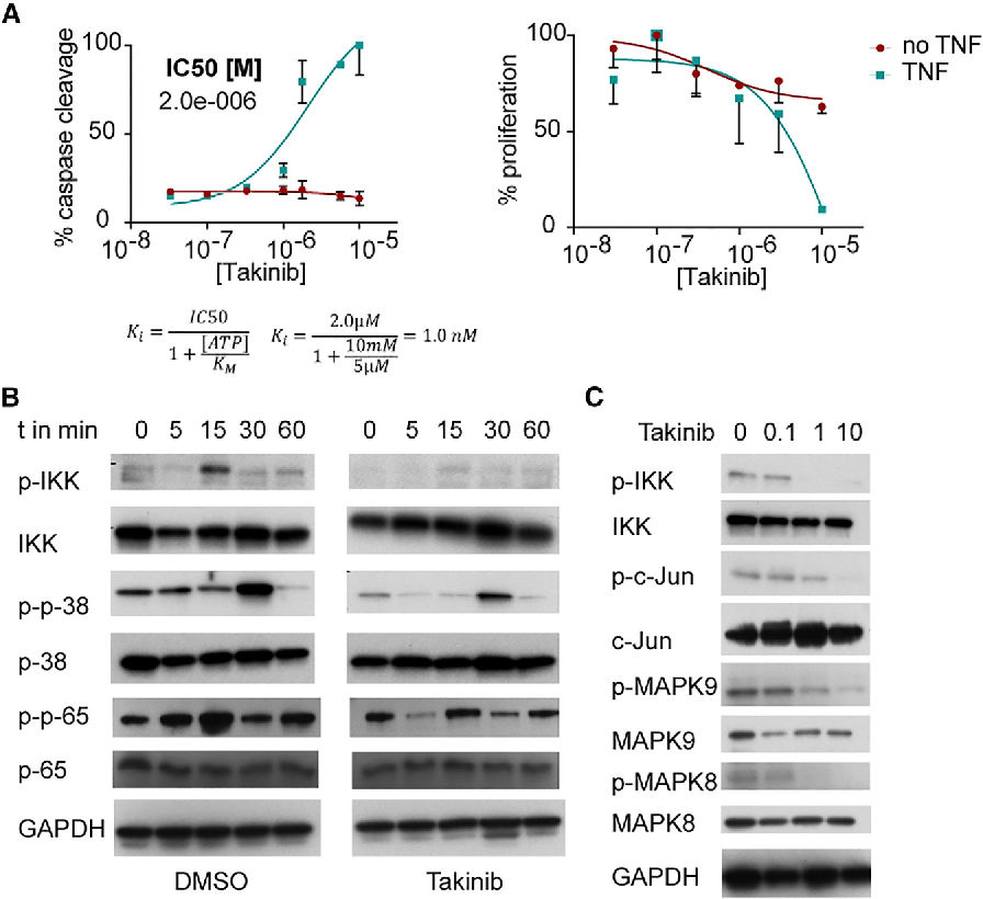
When the enzyme was activated with 5 μM ATP for 3 hr, the same V max was reached for 0, 10, 50, and 100 nM Takinib, and K M increased for these concentrations, which implies that Takinib is an ATP-competitive inhibitor if TAK1 is ATP activated.
Development of a High-Throughput Assay for Identifying

Using Genetically Engineered Kinases to Screen for Novel
c-Src Kinase Assay/Inhibitor Screening Kit ( Cat # KA0058 V.01 ) Introduction The Src family of non-receptor protein tyrosine kinases plays critical roles in a variety of
Note that most tyrosine kinases have Km values for ATP in the range 10-150 µM, so for kinetic experiments it is important to use saturating concentrations of ATP …
Abstract. Protein kinases catalyse the addition of phosphate groups to Ser/Thr and Tyr residues in cognate substrates and are mutated or hyperactive in a variety of diseases, making them important targets for rationally designed drugs.
Protein kinase B (PKB)/Akt is a member of the AGC family of serine/threonine kinases, named for the original members, protein kinase A, cGMP-dependent protein kinase, and protein kinase C. AGC kinases require phosphorylation of both a residue in the kinase domain (Thr308
phosphorylation using another method such as in vitro solution assay. Using ProtoArray® Human Protein Microarrays, we have typically observed a true positive rate of ~80% for serine-threonine protein kinases. A true positive signal is defined as a phosphorylation signal observed on a protein microarray that is validated as a substrate using an in vitro solution assay (page 29 for details
Identifying specific kinase substrates through engineered

Assay of protein kinases using radiolabeled ATP a
A Chemical Genetic Approach for the Identification of


Combining chemical genetics and proteomics to identify
Chitosan-assisted permeabilization of ATP–biotin for live
lok sabha seats state wise 2017 pdf – ADP TR-FRET Red Assay BellBrook Labs Kinase Assays
PLOS ONE A Homogeneous High-Throughput Assay for


ADP-Specific Sensors Enable Universal Assay of Protein
YouTube Embed: No video/playlist ID has been supplied
Considerations for the design and reporting of enzyme
Note that most tyrosine kinases have Km values for ATP in the range 10-150 µM, so for kinetic experiments it is important to use saturating concentrations of ATP …
A Generic Homogenous Method for Measuring Kinase and
Document Content PRESENCE OF CONTROLS: were included on every plate. EXPECTED OUTCOME: Active compounds result in decreasing readout signal. The compounds were assayed in multiple independent instances using an identical protocol; each instance is called a ‘test’.
Intended Use CaM kinase II Assay Kit is primarily designed
Several scintillation proximity assay (SPA) formats for HTS of protein kinases have also been reported that use common immobilization formats (e.g., biotin/streptavidin) to capture radiolabeled peptide reaction products on scintillant-containing surfaces 20, 22, 25, 26. Generally, however, the peptide substrate used in such assay formats must be optimized for each particular kinase and
Protein Tyrosine Kinase Assay Kit Non-Radioactive PTK101
In vitro Protein Kinase Assay Using Yeast Sch9 Bio-protocol
A Chemical Genetic Approach for the Identification of
LanthaScreen® TR-FRET tyrosine kinase and protein kinase C assay Dual wavelength detection of terbium and fluorescein using dedicated optic settings Strong and long-lasting donor signal minimizes background Rapid and reliable detection of PKC´s and tyrosine kinase activity Introduction Protein Kinase C (PKC) enzymes are a diverse family of enzymes that under specific signaling conditions
ADP-Specific Sensors Enable Universal Assay of Protein
Assaying Protein Kinase Activity with Radiolabeled ATP
The activity of Recombinant Human ERK1 (Catalog # 1879-KS) was assayed in the presence of 0.2 mM ATP, 0.2 mM myelin basic protein (MBP) peptide, and 0.2 μg CD39L2 in 50 μL at room temperature for 15 minutes. The slope of the curve corresponds to a specific activity of 482 pmol/min/μg using the conversion factor of 3500 pmol/OD and the coupling rate of 0.597.
Identification of Novel Substrates of MAP Kinase Cascades
CDK6/CyclinD3 Kinase Assay Promega Corporation
Transcreener® ADP² Assay Kits BellBrook Labs Kinase
In vitro activity assays (for purified kinases) of ATP to ADP can be monitored directly using spectroscopy. The products appear identical to the starting materials due to identical absorption features and extinction coefficients. Kinase assays: in buffer, in cell lysate, in vivo Depending on your application, you should consider whether you need an assay that can be performed only with
Homogenous Kinase Assay using ATPlite™ PerkinElmer
PKC-θ in vitro Kinase Activity Assay —BIO-PROTOCOL
For example, ,substrates are often phosphorylated by a combination of kinases, however, kinase assays have yet to be coupled to a read-out that can localize a phosphorylation event to a specific amino acid residue of a substrate and determine the extent of the phosphorylation.
c-Src Kinase Assay/Inhibitor Screening Kit is designed to